MRD and Biomarkers
Quantitation of immunoglobulin free light chains is impacted by glycosylated monoclonal proteins
P-188: Quantitation of immunoglobulin free light chains is impacted by glycosylated monoclonal proteins
Friday, September 27, 2024


Maria Alice Willrich, PhD
Associate Professor of Laboratory Medicine and Pathology
Mayo Clinic
Rochester, Minnesota, United States
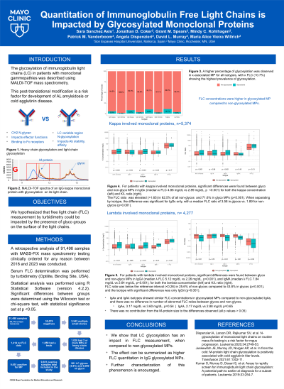
Introduction: The glycosylation of immunoglobulin light chains (LC) in patients with monoclonal gammopathies was described using MALDI-TOF mass spectrometry. This post-translational modification is a risk factor for development of AL amyloidosis or cold agglutinin disease. We hypothesized that free light chain (FLC) measurement by turbidimetry could be impacted by the presence of glyco groups on the surface of the light chains.
Methods: A retrospective analysis of 91,498 samples with MASS-FIX mass spectrometry testing clinically ordered for any reason between 2018 and 2023 was conducted. Serum FLC determination was performed by turbidimetry (Optilite, Binding Site, USA). Significant differences between groups were determined using the Wilcoxon test or chi-square test, with statistical significance set at p < 0.05. Statistical analysis was performed using R Statistical Software (version 4.2.2).
Results: From the 91,498 samples tested, only one measurement per patient was used (n=50,772). Out of those, 81% were not analyzed, as the samples were negative (n=35,078), had multiple small clones (n=2,542), more than one monoclonal protein (MP) or heavy chain disease (n=1,028), contained a therapeutic monoclonal antibody (n=1,059) or had no FLC data (n=1,414). The remainder of the samples (n=9,651) were positive for a single MP and included. 561 showed LC glycosylation (5.8%) and 9,090 did not. A higher percentage of glycosylation was observed in κ-associated MP for all isotypes, with κ FLC (10.7%) showing the highest prevalence of glycosylation. FLC concentrations were higher in glycosylated MP compared to non-glycosylated MPs. Significant differences were found between glycosylated and non-glycosylated MPs in IgGκ (median κ FLC 4.86 mg/dL vs 2.89 mg/dL, p < 0.001), IgGʎ (median λ FLC 5.12 mg/dL vs 2.26 mg/dL, p< 0.001), and IgMʎ (median λ FLC 7.84 mg/dL vs 2.64 mg/dL, p< 0.001). In contrast, IgAκ and IgAʎ isotypes showed similar FLC concentrations in glycosylated MPs compared to non-glycosylated IgAs (IgAκ, 3.17 mg/dL vs 3.60 mg/dL, p=0.54 and IgAλ, 2.17 mg/dL vs 2.88 mg/dL p=0.65). Among those with a κ-involved isotype, the FLC ratio was elevated ( >1.65) in 62.5% of all non-glycos and 71.6% in glyco MPs (p < 0.001). When separating by isotype, the difference was significant for IgGκ only, with a median FLC ratio of 3.58 in glycos vs. 1.89 for non-glycos (p < 0.001). Among those with λ-involved isotypes, FLC ratio was below the reference interval ( < 0.26) in 25.6% of non-glycos compared to 43.8% in glycos (p < 0.001), and the isotype with significant differences was only IgGʎ (p < 0.001). In IgAκ and ʎ isotypes, abnormal FLC ratio outside reference intervals was similar in glyco and non-glyco MPs.
Conclusions: We show that LC glycosylation has an impact in FLC measurement, when compared to non-glycosylated MPs. The effect can be summarized as higher FLC quantitation in IgG glycosylated MPs. Further characterization of this phenomenon is encouraged.